
Effect of Autologous Platelet-Rich Plasma (PRP) on Optical Quality and Visual Recovery after Trifocal Intraocular Lens Implantation
Purpose
To evaluate the effect of autologous platelet-rich plasma (PRP) eye drops on optical quality and visual acuity in eyes implanted with a diffractive trifocal intraocular lens (IOL).
Methods
In this retrospective cohort study of 128 eyes of 128 patients who underwent femtosecond laser-assisted cataract surgery with implantation of the CNWT Clareon PanOptix IOL (Alcon Inc.), patients were allocated into two groups: the PRP group (n = 62), which received PRP eye drops postoperatively, and the conventional treatment group (n = 66). Three weeks after surgery, optical parameters, including the objective scatter index (OSI), the modulation transfer function (MTF) cutoff and the Strehl ratio (SR), were measured with a double-pass system. Uncorrected distance visual acuity (UDVA) and corrected distance visual acuity (CDVA) at 4 m, as well as uncorrected near visual acuity (UNVA) at 40 cm, were recorded in logMAR units.
Results
The PRP group demonstrated superior optical quality in all measured parameters compared with the control group, with lower OSI (1.60 vs. 3.14), higher MTF cutoff (31.93 vs. 19.90 cycles/degree) and higher SR (0.15 vs. 0.12) (all P < 0.001). Visual acuity was also significantly better in the PRP group, with lower mean logMAR UDVA (0.05 vs. 0.10; P < 0.001), CDVA (0.01 vs. 0.05; P < 0.001) and UNVA (0.13 vs. 0.18; P = 0.007). Multivariate analyses confirmed that the use of PRP eye drops was independently associated with improvements in all three optical parameters: OSI (B = −1.488), MTF cutoff (B = 12.038) and SR (B = 0.039) (all P < 0.001).
Conclusions
The use of autologous PRP eye drops improves optical quality and visual acuity at all distances after diffractive trifocal intraocular lens implantation. PRP eye drops may be a beneficial adjunct in the postoperative management of patients undergoing this procedure.
Citation: Na KH, Lee BY, Cho HJ, Hyon JY (2026) Effect of autologous platelet-rich plasma on optical quality and visual outcomes after implantation of a diffractive trifocal intraocular lens. PLoS One 21(1): e0340880. https://doi.org/10.1371/journal.pone.0340880
Editor: Hyojin Kim, BaekSeok University, South Korea
Received: May 12, 2025; Accepted: December 29, 2025; Published: January 27, 2026
Copyright: © 2026 Na et al. This is an open access article distributed under the terms of the Creative Commons Attribution License , which permits unrestricted use, distribution, and reproduction in any medium, provided the original author and source are credited.
Data Availability: The research data may contain personal information and cannot be shared for ethical reasons. These restrictions were imposed by the Public Institutional Review Board designated by the Ministry of Health and Welfare to protect potentially identifying or sensitive patient information. However, if there is a reasonable request, it may be submitted to the Public Institutional Review Board ( irb@nibp.kr ) for review and approval.
Funding: The authors received no specific funding for this work.
Competing interests: The authors have declared that no competing interests exist.
Introduction
With the growing demand for presbyopia correction and reduced spectacle dependence after cataract surgery, multifocal intraocular lenses (IOLs) have been increasingly used [ 1 ]. Among them, diffractive trifocal IOLs have gained popularity because of their ability to provide satisfactory visual acuity at multiple distances [ 2–4 ]. However, concerns persist regarding the optical quality of multifocal IOLs compared with monofocal IOLs [ 5–7 ] . Considering that multifocal IOL implantation is typically performed in healthy individuals , without ocular or systemic comorbidities, and that these IOLs are more costly, achieving high-quality postoperative optical outcomes is of substantial clinical relevance, particularly to meet patients' high expectations [ 1 , 8 ] . Even without manifest ocular surface disease, subclinical tear film instability or subtle optical degradation can compromise visual satisfaction [ 8 ]. This underscores the relevance of interventions aimed at optimizing optical quality, even in clinically healthy eyes .
In recent years, there has been growing interest in blood-derived products, such as autologous serum (AS) or platelet-rich plasma (PRP), for the treatment of ocular surface diseases [ 9 ] . Autologous PRP has attracted particular attention because of its high concentration of platelet biological factors that promote ocular surface healing [ 10–12 ]. It has shown efficacy comparable or superior to AS in the treatment of dry eye syndrome (DES) and primary Sjögren's syndrome, with the added benefit of a shorter preparation time [ 13–15 ] . Furthermore, PRP has shown therapeutic potential in ocular surface disorders that do not respond to conventional therapies, such as indolent corneal ulcers and persistent corneal epithelial defects [ 11 , 12 , 16 , 17 ] .
Ocular surface irregularities, especially those associated with dry eye syndrome (DES), have been shown to negatively affect the optical performance of multifocal intraocular lenses (IOLs). A stable tear film is therefore essential for achieving optimal visual outcomes in eyes implanted with multifocal IOLs [ 8 ]. Previous studies have identified DES as a significant factor in patient dissatisfaction after presbyopia-correcting IOL implantation [ 18 , 19 ]. Based on this evidence, it is reasonable to hypothesize that PRP eye drops, by promoting ocular surface stabilization and tear film homeostasis, could improve optical and visual performance in eyes implanted with diffractive trifocal IOLs. To our knowledge, no previous study has investigated this hypothesis.
In this study, we aimed to evaluate the effects of autologous PRP eye drops on optical quality and visual outcomes after diffractive trifocal IOL implantation, comparing the conventional treatment (CT) group with the PRP group.
Methods
Study population
In this retrospective cohort study, we reviewed the medical records of consecutive patients who underwent femtosecond laser-assisted cataract surgery (FLACS) and implantation of the CNWT intraocular lens (IOL) (Clareon PanOptix; Alcon Inc., Fort Worth, TX, USA) between August 2022 and August 2023 at B&VIIT Eye Center, South Korea. This study followed the tenets of the Declaration of Helsinki and was approved by the Institutional Review Board (IRB) designated by the Ministry of Health and Welfare (Protocol number: P01-202405-01-018). The IRB waived the requirement for written informed consent due to the retrospective nature of the study and the use of anonymized data. The data were accessed on May 27, 2024, and the authors had no access to any identifiable participant information during or after data collection. All data were fully anonymized before access.
Group classification
Patients were divided into two groups: the CT group received the standard postoperative regimen of 0.5% moxifloxacin, 1% prednisolone and 0.45% ketorolac eye drops and preservative-free 0.18% hyaluronic acid (HA) artificial tears; the PRP group received PRP eye drops in addition to this regimen. Exclusion criteria included corneal astigmatism greater than 1.0 diopter (D), any ocular comorbidities other than cataract (such as corneal pathology, eyelid abnormalities, retinal or optic nerve diseases) and a history of ocular surgery. Patients with diabetes, autoimmune diseases (e.g., Sjögren's syndrome, rheumatoid arthritis), psychiatric disorders, thyroid diseases, allergic diseases, hematologic disorders, heart disease, acute or chronic infections, a history of cancer treatment, or who were on antiplatelet or anticoagulant therapy were also excluded. In addition, patients who did not attend the regular follow-up visits at 1 and 3 weeks after surgery were excluded.
Preoperative examinations
All patients underwent comprehensive ophthalmologic evaluations before surgery. Uncorrected distance visual acuity (UDVA), corrected distance visual acuity (CDVA) at 4 m and uncorrected near visual acuity (UNVA) at 40 cm were measured using Early Treatment Diabetic Retinopathy Study (ETDRS) charts and expressed in logMAR (logarithm of the minimum angle of resolution) units. Additional examinations included slit-lamp biomicroscopy, specular microscopy, corneal topography and tomography, dilated fundus examination and optical coherence tomography (OCT). Ocular biometry was performed using the IOLMaster 700 (Carl Zeiss Meditec AG, Jena, Germany), and intraocular lens (IOL) power for emmetropia was determined based on the Barrett Universal II formula.
Surgical techniques and postoperative management
All cataract surgeries were performed under topical anesthesia using the FLACS system with the LenSx system (Alcon Inc., Fort Worth, TX, USA) to create a 4.9 mm capsulorhexis and quadrant-pattern lens fragmentation. Phacoemulsification, irrigation, aspiration and polishing were then performed with the CENTURION Vision system (Alcon Inc., Fort Worth, TX, USA). The IOL was implanted in the capsular bag, and all incisions were sealed by stromal hydration without sutures. All surgeries were uneventful, and no intraoperative complications, such as posterior capsule rupture, zonular dehiscence or significant intraoperative miosis, occurred in any case. Postoperatively, patients instilled 0.5% moxifloxacin and 1% prednisolone eye drops four times a day, 0.45% ketorolac eye drops twice a day and 0.18% hyaluronic acid artificial tears six times a day. Follow-up visits were scheduled for 1 and 3 weeks after surgery.
Preparation of autologous platelet-rich plasma eye drops
At the 1-week postoperative visit, PRP eye drops were prepared for the PRP group using the 3E-PRP kit (Pervice Co., Ltd., South Korea) according to the manufacturer's instructions [ 20 ]. The manufacturer reports that the final platelet concentration obtained with this method is approximately 9.3 to 12 times higher than the baseline level in whole blood [ 20 ]. We collected 8.5 mL of venous blood into sterile 10 mL tubes containing 1.5 mL of acid citrate dextrose solution A anticoagulant. After gentle homogenization, the samples were centrifuged at 3,000 rpm for 5 minutes to obtain three layers: (1) an upper plasma layer, (2) an intermediate platelet-rich layer and (3) a lower red blood cell layer. Using the supplied connector, we aspirated the supernatant plasma and buffy coat into separate syringes, combining them to form the PRP, and divided the final product into 2 vials, each containing approximately 2 mL. Patients were instructed to instill PRP drops four times a day for 2 weeks, starting 1 week after surgery; opened vials were stored at 4 °C and unopened vials at −20 °C.
Measurement of optical quality parameters and other postoperative evaluations.
Optical quality was evaluated at the 3-week postoperative visit using the double-pass optical quality analysis system (OQAS II; Visiometrics, Spain), which assesses the combined effect of ocular aberrations and intraocular scattering [ 21–23 ] . This system captures images of a 780 nm point-source object reflected on the retina and directly calculates the modulation transfer function (MTF) from the acquired double-pass retinal image using the Fourier transform. It also provides MTF-related parameters, such as the MTF cutoff and the Strehl ratio (SR). The MTF cutoff is the highest spatial frequency detectable by the eye, with higher values indicating better optical quality. SR is defined as the ratio between the area under the MTF curve of the measured optical system and that of an aberration-free system, with higher SR values corresponding to better optical performance [ 22 , 24 ]. Additionally, the system provides the objective scatter index (OSI), which quantifies intraocular light scattering. OSI is defined as the ratio between the light intensity at a peripheral location and the central peak in the double-pass image, with higher OSI values indicating greater intraocular scattering and visual disturbance [ 22 , 23 , 25 ]. All measurements were performed under mesopic conditions with a 4.0 mm artificial pupil by experienced technicians.
Non-invasive keratograph tear film break-up time (NIKBUT) was measured at the 1- and 3-week postoperative visits using the Keratograph 5M (Oculus, Wetzlar, Germany) [ 26 , 27 ]. Both the time of the first tear film break-up (NIKBUT-first) and the average time of all tear film break-ups (NIKBUT-avg) were recorded.
Uncorrected (UDVA) and corrected (CDVA) distance visual acuity at 4 m and uncorrected near visual acuity (UNVA) at 40 cm were measured at the 3-week postoperative evaluation. In addition, total corneal higher-order aberrations (HOAs) at a 6 mm diameter, spherical aberration (SA) at a 4 mm diameter and the degree of intraocular lens (IOL) decentration and tilt were assessed using anterior segment swept-source optical coherence tomography (CASIA2; Tomey, Japan).
Statistical analysis
Statistical analyses were performed using SPSS for Windows (version 28.0; SPSS Inc., Chicago, Illinois, USA) and R software (version 4.4.0; R Foundation for Statistical Computing, Vienna, Austria). Continuous variables were compared using the independent-samples Student's t test , and categorical variables were analyzed using the chi-square test. Jitter plots were used to visually represent the distribution of optical quality parameters. Univariate and multivariate linear regression analyses were conducted to identify factors associated with OSI, MTF cutoff and SR. Variables with a p value below 0.05 in the univariate analysis were included in the multivariate model to assess their independent effects. Multicollinearity was assessed using the variance inflation factor (VIF), with a VIF greater than 10 considered indicative of significant collinearity [ 28 ]. A p value below 0.05 was considered statistically significant.
Results
Baseline characteristics
Of the 148 patients initially enrolled, 20 were excluded for the following reasons: previous laser vision correction surgery (n = 4), diabetes (n = 6), rheumatoid arthritis (n = 1), Sjögren's syndrome (n = 1), hypothyroidism (n = 1), current use of antiplatelet agents (n = 4) and hepatitis B virus carrier status (n = 3). Consequently, 128 eyes of 128 patients were included in the final analysis, with 62 patients allocated to the PRP group and 66 patients to the CT group.
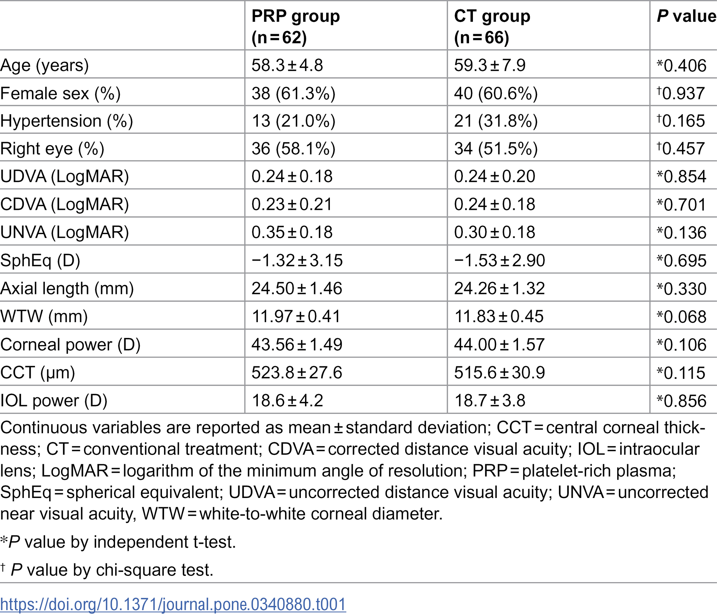
The mean age in both groups was in the late 50s, and approximately 60% of patients were female. No significant differences were observed between the two groups in preoperative visual acuity, spherical equivalent (SE), axial length, white-to-white corneal diameter, corneal power, central corneal thickness (CCT) and IOL power ( Table 1 ).
Optical quality parameters
The distribution of OSI, MTF cutoff and SR values measured 3 weeks after surgery is shown in Figure 1. Mean OSI was significantly lower in the PRP group than in the CT group (1.60 ± 0.80 vs. 3.14 ± 1.91, P < 0.001; Figure 1A ). Mean MTF cutoff was significantly higher in the PRP group compared with the CT group (31.93 ± 10.52 vs. 19.90 ± 9.93 cycles per degree, P < 0.001; Figure 1B ). Likewise, mean SR was significantly higher in the PRP group than in the CT group (0.15 ± 0.06 vs. 0.12 ± 0.04, P < 0.001; Figure 1C ).
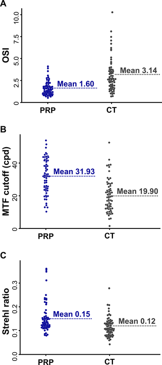
Figure 1. Distribution of the objective scatter index (OSI) (A), modulation transfer function (MTF) cutoff (B) and Strehl ratio (SR) (C) in eyes implanted with the CNWT intraocular lens, stratified by postoperative use of autologous platelet-rich plasma (PRP) eye drops.
The PRP group showed lower OSI and higher MTF cutoff and SR values, indicating superior optical performance compared with the conventional treatment (CT) group. cpd = cycles per degree.
https://doi.org/10.1371/journal.pone.0340880.g001
Non-invasive keratograph break-up time
At the 1-week postoperative evaluation, NIKBUT-first was 8.02 ± 5.94 seconds in the PRP group and 9.45 ± 6.80 seconds in the CT group, with no significant difference ( P = 0.210). At 3 weeks postoperatively, NIKBUT-first increased to 10.37 ± 6.60 seconds in the PRP group and decreased to 6.57 ± 4.90 seconds in the CT group, with a significant difference between groups ( P < 0.001; Fig. 2A ). Likewise, NIKBUT-avg was 10.74 ± 5.69 seconds in the PRP group and 12.56 ± 5.99 seconds in the CT group at 1 week postoperatively ( P = 0.080). After 3 weeks, NIKBUT-avg increased to 12.72 ± 5.96 seconds in the PRP group, while it decreased to 9.62 ± 5.14 seconds in the CT group ( P = 0.002, Fig 2B ).
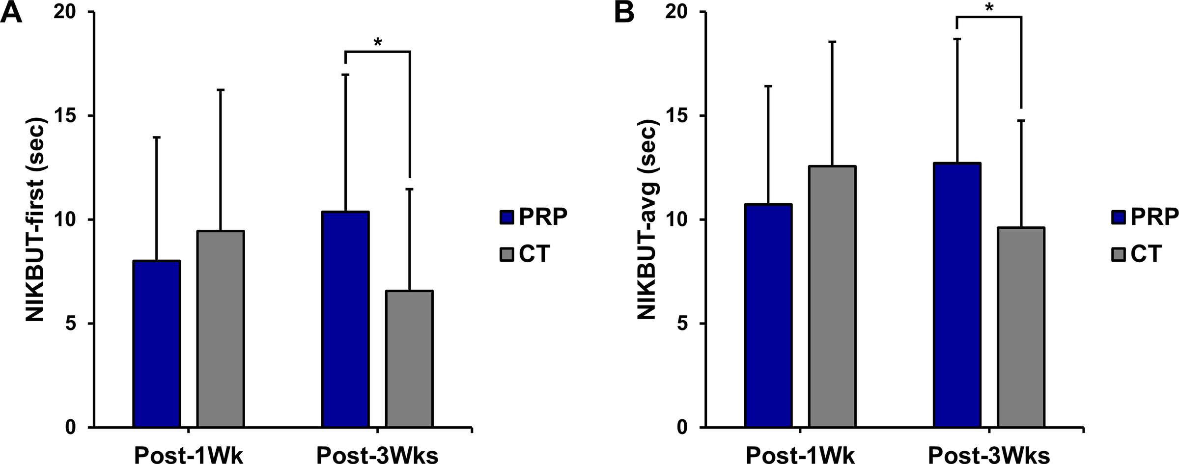
Figure 2. Tear film break-up time measured by non-invasive keratography (NIKBUT) 1 week and 3 weeks after surgery.
CT = conventional treatment group; NIKBUT-avg = average NIKBUT of all tear film break-ups; NIKBUT-first = NIKBUT of the first tear film break-up; Post-1wk = 1 week after surgery; Post-3wk = 3 weeks after surgery; PRP = platelet-rich plasma group; s = seconds. The asterisk [*] indicates P < 0,05.
https://doi.org/10.1371/journal.pone.0340880.g002
Visual outcomes and intraocular lens position
The PRP group demonstrated superior visual acuity compared with the CT group. Mean logMAR UDVA (uncorrected distance visual acuity) was 0.05 ± 0.06 in the PRP group and 0.10 ± 0.10 in the CT group ( p < 0.001). Mean logMAR CDVA (corrected distance visual acuity) was 0.01 ± 0.03 in the PRP group and 0.05 ± 0.07 in the CT group ( p < 0.001). For UNVA, mean values were 0.13 ± 0.10 in the PRP group and 0.18 ± 0.13 in the CT group ( p = 0.007). No significant differences were observed between the two groups in terms of spherical equivalent (SphEq), corneal higher-order aberrations (HOAs), spherical aberration (SA) or the degree of IOL decentration and tilt ( Table 2 ).
Table 2. Postoperative visual outcomes and intraocular lens position.
https://doi.org/10.1371/journal.pone.0340880.t002
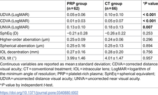
Table 2. Postoperative visual outcomes and intraocular lens position.
https://doi.org/10.1371/journal.pone.0340880.t002
Factors associated with optical quality parameters
In the univariate analysis for OSI, the use of PRP eye drops and postoperative SphEq were identified as significant factors ( P < 0.05) and included in the multivariate analysis. In the multivariate regression analysis, only the use of PRP eye drops remained significantly associated with lower OSI values (B = –1.488 ± 0.261, P < 0.001; Table 3 ). Regarding MTF cutoff and SR, the use of PRP eye drops was the only significant factor associated with higher MTF cutoff (B = 12.038 ± 1.808) and SR (B = 0.039 ± 0.009) values ( P < 0.001), thus making additional multivariate analyses unnecessary ( Table 3 ).
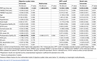
Table 3. Univariate and multivariate linear regression analysis for optical quality parameters.
https://doi.org/10.1371/journal.pone.0340880.t003
Associations between optical quality parameters and postoperative visual acuity
Given the superior optical quality and better visual acuity outcomes observed in the PRP group, additional analyses were conducted to evaluate the relationship between optical quality parameters and postoperative visual acuity. Multivariate analysis revealed that improved optical quality was associated with better visual acuity outcomes. Specifically, lower OSI values, indicating better optical quality, were associated with better UDVA (B = 0.012 ± 0.004, P = 0.005), while higher MTF cutoff (B = −0.002 ± 0.001, P = 0.011) and SR (B = −0.251 ± 0.125, P = 0.047) values, also indicating better optical quality, were associated with better UDVA. Regarding CDVA, only OSI showed a significant association (B = 0.007 ± 0.003, P = 0.028). For UNVA, both OSI (B = 0.019 ± 0.006, P = 0.002) and MTF cutoff (B = −0.002 ± 0.001, P = 0.010) showed significant correlations ( Table 4 ).
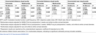
Table 4. Univariate and multivariate linear regression analysis for postoperative visual acuity.
https://doi.org/10.1371/journal.pone.0340880.t004
Discussion
To our knowledge, this is the first study to investigate the effect of autologous PRP eye drops on optical quality and visual outcomes after diffractive trifocal IOL implantation.
This study evaluated outcomes during the early postoperative period, 3 weeks after surgery. A functional magnetic resonance imaging study showed that patients implanted with diffractive IOLs had increased cortical activity 3 weeks after surgery [ 29 ], indicating the onset of the neuroadaptation process to multifocal IOLs.
Considering the crucial role of the ocular surface and tear film in maintaining visual quality [ 30 , 31 ], ocular surface irregularities in this early phase may negatively affect neuroadaptation and, consequently, impact long-term patient satisfaction [ 19 , 32 , 33 ]. Therefore, efforts to stabilize the ocular surface and optimize optical quality during the early postoperative period are particularly relevant after diffractive trifocal IOL implantation, especially in a preoperatively healthy population with high expectations. Future studies with longer follow-up periods are needed to evaluate the sustained effects of PRP eye drops on optical quality and visual outcomes beyond the early postoperative phase.
To minimize the likelihood that postoperative differences in optical quality were influenced by pre-existing ocular surface abnormalities, the functional baseline was defined as the 1-week postoperative visit, the time point immediately before the start of PRP treatment in the PRP group. All patients underwent comprehensive preoperative screening, and individuals with ocular surface disease or systemic conditions known to affect tear film stability (e.g., Sjögren's syndrome, rheumatoid arthritis, thyroid dysfunction or diabetes) were excluded [ 34 ]. Importantly, at this 1-week baseline visit, NIKBUT values did not differ significantly between the PRP and control (CT) groups, indicating comparable tear film stability at the functional baseline. Although corneal staining was not assessed, which is a limitation of the study, the similarity in NIKBUT supports the assumption that both groups started from a comparable level of ocular surface integrity and dryness before the initiation of PRP therapy [ 26 , 27 ].
In this study, optical quality was evaluated using OQAS parameters, including OSI, MTF cutoff and SR, based on previous work demonstrating the usefulness of these measures for objectively assessing optical quality in pseudophakic eyes [ 35–37 ] . Although direct comparisons are challenging, the optical quality parameters observed in the PRP group were comparable to those reported for monofocal IOLs in previous studies and even to those of individuals without lens opacity [ 22 ]. To better characterize the independent association between the use of PRP eye drops and optical quality, we included covariates such as IOL decentration [ 38 ], tilt [ 39 ] and tear film break-up time [ 31 , 40 ] in multivariate analyses, since these factors are known to influence optical quality after multifocal IOL implantation. This approach allowed a more precise characterization of the relationship between the use of PRP eye drops and optical quality, an area that has not been comprehensively addressed in previous studies [ 35–37 ].
Platelets are essential reservoirs of growth factors, such as platelet-derived growth factor and epidermal growth factor, which are stored predominantly in their α-granules [ 9 , 10 ]. These growth factors promote tissue regeneration and healing, contributing to the broad therapeutic potential of PRP products. In ophthalmology, PRP has shown promising effects in the treatment of ocular surface diseases refractory to conventional therapies. The ocular surface restorative effects of PRP observed in previous studies are also reflected in our findings.
Although the platelet concentration of the PRP prepared in this study was not directly analyzed, the manufacturer's technical brochure indicates that the 3E-PRP system yields a final platelet concentration approximately 9.3 to 12 times higher than the baseline whole blood level [ 20 ]. PRP contains a variety of bioactive components beyond platelets, including multiple growth factors that play critical roles in tissue repair and ocular surface healing [ 10 – 12 ]. Future studies incorporating detailed compositional or proteomic analyses, including quantification of platelet concentration and characterization of growth factors and other bioactive molecule profiles, will be valuable for elucidating the components strongly associated with clinical efficacy and for establishing standardized preparation and dosing protocols for ophthalmic PRP applications.
The PRP group showed a significant improvement in NIKBUT 3 weeks after surgery, whereas previous reports, consistent with the findings in the CT group, have shown a decrease in tear film break-up time after cataract surgery [ 41 , 42 ]. Despite this improvement in NIKBUT, no direct association with optical quality parameters was found in this study. This may be because NIKBUT does not fully capture the spatial variability in tear film stability. The impact on optical quality in eyes with diffractive trifocal IOLs may vary depending on whether tear film disturbances occur centrally or peripherally, a distinction not accounted for by NIKBUT. In addition, factors such as corneal morphology and transparency also influence optical quality [ 43 , 44 ]. Therefore, further studies incorporating more detailed tear film assessments and corneal evaluations are needed to elucidate the mechanisms underlying the improvements observed in the PRP group.
Multivariate analyses revealed significant correlations between OQAS parameters and visual acuity outcomes. This is consistent with the observation that the PRP group showed superior optical quality and visual acuity compared with the CT group. Previous studies have also demonstrated significant associations between OQAS parameters and visual acuity, supporting the findings of the present investigation [ 45 – 47 ].
All patients in this study underwent cataract surgery with a standardized femtosecond laser capsulotomy size and a uniform quadrant nuclear fragmentation pattern. This standardization is important, as capsulotomy size and shape can affect IOL position [ 48 ], potentially influencing optical quality outcomes. Such procedural uniformity enhances the internal validity of our study.
Although the present study consecutively included all eligible patients who underwent femtosecond laser-assisted cataract surgery (FLACS) with CNWT intraocular lens (IOL) implantation during the study period, the relatively strict exclusion criteria limit the external validity of the results. These criteria reflect the narrow indication for multifocal IOL implantation, which is generally limited to eyes without ocular or systemic comorbidities that could negatively affect postoperative visual performance [ 1 , 8 ]. In addition, exclusions relevant to the safe or appropriate use of autologous platelet-rich plasma (PRP), such as current use of antiplatelet agents and infectious status, including hepatitis B carrier status, were applied equally to both groups to maintain comparability and reduce potential confounders [ 49 , 50 ]. As a result, the study population represents a highly selected subset of patients undergoing cataract surgery; however, this approach was necessary to maintain cohort homogeneity and minimize confounding in the assessment of ocular surface and optical quality outcomes in the immediate postoperative period.
It is crucial to acknowledge the following limitations when interpreting our findings. First, the retrospective, non-randomized design introduces inherent constraints. Although baseline characteristics were similar between the PRP and CT groups and multivariate analyses were performed to control for confounders, unmeasured variables, such as socioeconomic status, lifestyle factors and adherence to postoperative regimens, may have influenced treatment allocation and outcomes. Future randomized controlled trials are needed to validate these findings. Second, corneal staining was not assessed. Although NIKBUT provides a non-invasive measure of tear film stability and avoids dye-induced reflex tearing, corneal staining could offer additional information on epithelial integrity and its impact on optical quality. Third, PRP eye drops were used together with antibiotics, steroid eye drops and HA artificial tears; the observed improvements should therefore be considered additive rather than attributable exclusively to PRP. Fourth, the follow-up duration was relatively short, which may not fully capture the long-term stability of ocular surface recovery or the persistence of optical quality improvements. Long-term studies are needed to determine whether the early benefits of PRP are maintained over time. Fifth, patient-reported outcomes were not collected. Instruments such as the Ocular Surface Disease Index [ 51 ] or visual quality questionnaires [ 52 ] would provide complementary subjective information and help contextualize the clinical relevance of the observed optical improvements. Future studies integrating subjective and objective measures will enhance the generalizability and translational value of these findings.
In conclusion, autologous PRP eye drops improve optical quality and visual outcomes after diffractive trifocal IOL implantation. Further studies, including detailed corneal surface assessments and longer follow-up periods, are needed to provide deeper insight into the effects of PRP on optical and visual performance in eyes with diffractive trifocal IOLs.
References
1.Rampat R, Gatinel D. Multifocal and Extended Depth-of-Focus Intraocular Lenses in 2020. Ophthalmology. 2021;128(11):e164–85. pmid:32980397
2.Kohnen T, Herzog M, Hemkeppler E, Schönbrunn S, De Lorenzo N, Petermann K, et al. Visual Performance of a Quadrifocal (Trifocal) Intraocular Lens Following Removal of the Crystalline Lens. Am J Ophthalmol. 2017;184:52–62. pmid:28923587
3.Bilbao-Calabuig R, Llovet-Rausell A, Ortega-Usobiaga J, Martínez-Del-Pozo M, Mayordomo-Cerdá F, Segura-Albentosa C, et al. Visual Outcomes Following Bilateral Implantation of Two Diffractive Trifocal Intraocular Lenses in 10 084 Eyes. Am J Ophthalmol. 2017;179:55–66. pmid:28456547
4.Brenner LF, Nistad K, Schonbeck U. Rethinking presbyopia: results of bilateral refractive lens exchange with trifocal intraocular lenses in 17,603 patients. Br J Ophthalmol. 2023;107(7):912–9. pmid:35110276
5.Na KS, Kim SJ, Nam G, Ha M, Whang WJ, Kim EC, et al. A Novel Intraocular Lens Simulator That Enables Patients to Experience the World Through Multifocal Intraocular Lenses Before Surgery. Transl Vis Sci Technol. 2022;11(3):14. pmid:35275206
6.Kim EC, Na KS, Kim HS, Hwang HS. How does the world appear to patients with multifocal intraocular lenses?: a mobile model eye experiment. BMC Ophthalmol. 2020;20(1):180. pmid:32375711
7.Kim EC, Cho SY, Kang JE, Nam G, Yoon YC, Whang WJ, et al. Comparative Analysis of the Optical Quality of Monofocal, Enhanced Monofocal, Multifocal, and Extended Depth of Focus Intraocular Lenses: A Mobile Model Eye Study. Transl Vis Sci Technol. 2023;12(7):5. pmid:37405796
8.Alio JL, Plaza-Puche AB, Férnandez-Buenaga R, Pikkel J, Maldonado M. Multifocal intraocular lenses: An overview. Surv Ophthalmol. 2017;62(5):611–34. pmid:28366683
9.Anitua E, Muruzabal F, Tayebba A, Riestra A, Perez VL, Merayo-Lloves J, et al. Autologous serum and plasma rich in growth factors in ophthalmology: preclinical and clinical studies. Acta Ophthalmol. 2015;93(8):e605-14. pmid:25832910
10.Nurden AT. Platelets, inflammation and tissue regeneration. Thromb Haemost. 2011;105 Suppl 1:S13-33. pmid:21479340
11.Kim KM, Shin YT, Kim HK. Effect of autologous platelet-rich plasma on persistent corneal epithelial defect after infectious keratitis. Jpn J Ophthalmol. 2012;56(6):544–50. pmid:22972393
12.Alio JL, Rodriguez AE, Wróbel-Dudzińska D. Eye platelet-rich plasma in the treatment of ocular surface disorders. Curr Opin Ophthalmol. 2015;26(4):325–32. pmid:26058033
13.Metheetrairut C, Ngowyutagon P, Tunganuntarat A, Khowawisetsut L, Kittisares K, Prabhasawat P. Comparison of epitheliotrophic factors in platelet-rich plasma versus autologous serum and their treatment efficacy in dry eye disease. Sci Rep. 2022;12(1):8906. pmid:35618742
14.Kang MJ, Lee JH, Hwang J, Chung SH. Efficacy and safety of platelet-rich plasma and autologous-serum eye drops for dry eye in primary Sjögren's syndrome: a randomized trial. Sci Rep. 2023;13(1):19279. pmid:37935760
15.Wróbel-Dudzińska D, Przekora A, Kazimierczak P, Ćwiklińska-Haszcz A, Kosior-Jarecka E, Żarnowski T. The Comparison between the Composition of 100% Autologous Serum and 100% Platelet-Rich Plasma Eye Drops and Their Impact on the Treatment Effectiveness of Dry Eye Disease in Primary Sjogren Syndrome. J Clin Med. 2023;12(9):3126. pmid:37176566
16.Alio JL, Abad M, Artola A, Rodriguez-Prats JL, Pastor S, Ruiz-Colecha J. Use of autologous platelet-rich plasma in the treatment of dormant corneal ulcers. Ophthalmology. 2007;114(7):1286-1293.e1. pmid:17324465
17.Alio JL, Rodriguez AE, Abdelghany AA, Oliveira RF. Autologous Platelet-Rich Plasma Eye Drops for the Treatment of Post-LASIK Chronic Ocular Surface Syndrome. J Ophthalmol. 2017;2017:2457620. pmid:29379652
18.Gibbons A, Ali TK, Waren DP, Donaldson KE. Causes and correction of dissatisfaction after implantation of presbyopia-correcting intraocular lenses. Clin Ophthalmol. 2016;10:1965–70. pmid:27784985
19.Woodward MA, Randleman JB, Stulting RD. Dissatisfaction after multifocal intraocular lens implantation. J Cataract Refract Surg. 2009;35(6):992–7. pmid:19465282
20.Pervice Co., Ltd. 3E-PRP system technical brochure. Includes the manufacturer-provided preparation protocol and platelet concentration data (based on platelet count testing by Green Cross Medical Foundation). https://oyasama.es/wp-content/uploads/2022/02/3E-PRP-brochure-NEW.pdf
21.Güell JL, Pujol J, Arjona M, Diaz-Douton F, Artal P. Optical Quality Analysis System; Instrument for objective clinical evaluation of ocular optical quality. J Cataract Refract Surg. 2004;30(7):1598–9. pmid:15210251
22.Saad A, Saab M, Gatinel D. Repeatability of measurements with a double-pass system. J Cataract Refract Surg. 2010;36(1):28–33. pmid:20117702
23.Cabot F, Saad A, McAlinden C, Haddad NM, Grise-Dulac A, Gatinel D. Objective assessment of crystalline lens opacity level by measuring ocular light scattering with a double-pass system. Am J Ophthalmol. 2013;155(4):629–35, 635.e1-2. pmid:23317652
24.Díaz-Doutón F, Benito A, Pujol J, Arjona M, Güell JL, Artal P. Comparison of the retinal image quality with a Hartmann-Shack wavefront sensor and a double-pass instrument. Invest Ophthalmol Vis Sci. 2006;47(4):1710–6. pmid:16565413
25.Artal P, Benito A, Pérez GM, Alcón E, De Casas A, Pujol J, et al. An objective scatter index based on double-pass retinal images of a point source to classify cataracts. PLoS One. 2011;6(2):e16823. pmid:21326868
26.Hong J, Sun X, Wei A, Cui X, Li Y, Qian T, et al. Assessment of tear film stability in dry eye with a newly developed keratograph. Cornea. 2013;32(5):716–21. pmid:23132457
27.Tian L, Qu JH, Zhang XY, Sun XG. Repeatability and Reproducibility of Noninvasive Keratograph 5M Measurements in Patients with Dry Eye Disease. J Ophthalmol. 2016;2016:8013621. pmid:27190639
28.Dormann CF, Elith J, Bacher S, Buchmann C, Carl G, Carré G, et al. Collinearity: a review of methods to deal with it and a simulation study evaluating their performance. Ecography. 2012;36(1):27–46.
29.Rosa AM, Miranda ÂC, Patrício MM, McAlinden C, Silva FL, Castelo-Branco M, et al. Functional magnetic resonance imaging to assess neuroadaptation to multifocal intraocular lenses. J Cataract Refract Surg. 2017;43(10):1287–96. pmid:29120714
30.Koh S. Mechanisms of Visual Disturbance in Dry Eye. Cornea. 2016;35 Suppl 1:S83–8. pmid:27583799
31.Herbaut A, Liang H, Rabut G, Trinh L, Kessal K, Baudouin C, et al. Impact of Dry Eye Disease on Vision Quality: An Optical Quality Analysis System Study. Transl Vis Sci Technol. 2018;7(4):5. pmid:30009091
32.Alió JL, Pikkel J. Multifocal Intraocular Lenses: Neuroadaptation. Alió JL, Pikkel J. Multifocal Intraocular Lenses. Springer. 2019. 53–60.
33.de Vries NE, Webers CAB, Touwslager WRH, Bauer NJC, de Brabander J, Berendschot TT, et al. Dissatisfaction after implantation of multifocal intraocular lenses. J Cataract Refract Surg. 2011;37(5):859–65. pmid:21397457
34.Qian L, Wei W. Identified risk factors for dry eye syndrome: A systematic review and meta-analysis. PLoS One. 2022;17(8):e0271267. pmid:35984830
35.Jiménez R, Valero A, Fernández J, Anera RG, Jiménez JR. Optical quality and visual performance after cataract surgery with biaxial microincision intraocular lens implantation. J Cataract Refract Surg. 2016;42(7):1022–8. pmid:27492101
36.Liao X, Li JY, Tan QQ, Tian J, Lin J, Lan CJ. Comparison of visual quality after implantation of A1-UV and SN60WF aspheric intraocular lenses. Int J Ophthalmol. 2020;13(11):1727–32. pmid:33215002
37.Lee H, Lee K, Ahn JM, Kim EK, Sgrignoli B, Kim TI. Double-pass system assessing the optical quality of pseudophakic eyes. Optom Vis Sci. 2014;91(4):437–43. pmid:24492759
38.Ortiz C, Esteve-Taboada JJ, Belda-Salmerón L, Monsálvez-Romín D, Domínguez-Vicent A. Effect of Decentration on the Optical Quality of Two Intraocular Lenses. Optom Vis Sci. 2016;93(12):1552–9. pmid:27776082
39.Oshika T, Sugita G, Miyata K, Tokunaga T, Samejima T, Okamoto C, et al. Influence of tilt and decentration of scleral-sutured intraocular lens on ocular higher-order wavefront aberration. Br J Ophthalmol. 2007;91(2):185–8. pmid:16914469
40.Gao Y, Liu R, Liu Y, Ma B, Yang T, Hu C, et al. Optical quality in patients with dry eye before and after treatment. Clin Exp Optom. 2021;104(1):101–6. pmid:32618024
41.Kasetsuwan N, Satitpitakul V, Changul T, Jariyakosol S. Incidence and pattern of dry eye after cataract surgery. PLoS One. 2013;8(11):e78657. pmid:24265705
42.Shimabukuro M, Maeda N, Koh S, Abe K, Kobayashi R, Nishida K. Effects of cataract surgery on symptoms and findings of dry eye in subjects with and without preexisting dry eye. Jpn J Ophthalmol. 2020;64(4):429–36. pmid:32495157
43.McCally RL, Farrell RA. Light scattering from cornea and corneal transparency. Noninvasive Diagnostic Techniques in Ophthalmology. Springer New York. 1990. 189–210. https://doi.org/10.1007/978-1-4613-8896-8_12
44.Meek KM. Corneal collagen: its role in maintaining corneal shape and transparency. Biophys Rev. 2009;1(2):83–93. pmid:28509987
45.Martinez-Roda JA, Vilaseca M, Ondategui JC, Almudi L, Asaad M, Mateos-Pena L, et al. Double-pass technique and compensation-comparison method in eyes with cataract. J Cataract Refract Surg. 2016; 42:1461–9. doi: https://doi.org/10.1016/j.jcrs.2016.08.015 . pmid:27839601
46.Martínez-Roda JA, Vilaseca M, Ondategui JC, Aguirre M, Pujol J. Effects of aging on optical quality and visual function. Clin Exp Optom. 2016;99(6):518–25. pmid:27452417
47.Li Y, Jin L, Wu M, Huang Y. Value of subjective visual quality assessment in early cataract surgical indications based on objective scatter index values. Front Med (Lausanne). 2022;9:1075693. pmid:36582278
48.Rossi T, Ceccacci A, Testa G, Ruggiero A, Bonora N, D'Agostino I, et al. Influence of anterior capsulorhexis shape, centration, size, and location on intraocular lens position: finite element model. J Cataract Refract Surg. 2022;48(2):222–9. pmid:34117178
49.Wu PI-K, Diaz R, Borg-Stein J. Platelet-Rich Plasma. Phys Med Rehabil Clin N Am. 2016;27(4):825–53. pmid:27788903
50.Eymard F, Louati K, Noel É, Abouqal R, Adam P, Allali F, et al. Indications and contraindications for platelet-rich plasma injections in musculoskeletal diseases in cases of infectious, oncological and hematological comorbidities: a 2025 formal consensus from GRIIP (Groupe de Recherche International sur les Injections de Plaquettes). Knee Surg Sports Traumatol Arthrosc. 2025;33(6):2293–306. pmid:40260684
51.Pult H, Wolffsohn JS. The development and evaluation of the new Ocular Surface Disease Index-6. Ocul Surf. 2019;17(4):817–21. pmid:31442595
52.McAlinden C, Pesudovs K, Moore JE. The development of an instrument to measure quality of vision: the Quality of Vision (QoV) questionnaire. Invest Ophthalmol Vis Sci. 2010;51(11):5537–45. pmid:20505205


Comments