
Safety of umbilical cord blood platelet-rich plasma applied to the subretinal space in patients with geographic atrophy associated with AMD
Authors
Stanislao Rizzo; Maria Cristina Savastano; Benedetto Falsini; Patrizio Bernardinelli; Francesco Boselli; Umberto De Vico; Matteo Mario Carlà; Federico Giannuzzi; Claudia Fossataro; Gloria Gambini; Emanuele Crincoli; Silvia Ferrara; Matteo Ripa; Rafael Killian; Clara Rizzo; Caterina Giovanna Valentini; Nicoletta Orlando; Giorgio Placidi; Luciana Teofili; Alfonso Savastano.
DOI: 10.1016/j.xops.2024.100476Publication type: open-access scientific article.
Purpose
The main objective of the study was to analyze the safety of the subretinal application of platelet-rich plasma obtained from umbilical cord blood, known as CB-PRP.
In addition to safety, the researchers also investigated whether the procedure could produce any preliminary sign of benefit in eyes with geographic atrophy related to the dry form of age-related macular degeneration.
Design
The work was part of the Si.Cord Study, which began in January 2021 and was conducted with 12 months of clinical follow-up.
It was a clinical trial that was:
interventional;
open-label;
single-center;
non-randomized;
with sequential allocation.
The research was carried out at the Fondazione Policlinico Universitario Agostino Gemelli IRCCS, in Rome, Italy.
The study was registered on ClinicalTrials.gov under the code NCT04636853.
Participants
A total of 13 patients were included, totaling 26 eyes, all with bilateral geographic atrophy associated with age-related macular degeneration.
In each patient, the eye with the more advanced involvement was chosen to receive the treatment. The fellow eye remained untreated and was used as a control for comparison.
All participants completed the 12-month follow-up period.
Intervention
The 13 selected eyes underwent 23 G vitrectomy.
After the vitrectomy, a subretinal injection of CB-PRP was performed using a 41-gauge needle.
The product was prepared from umbilical cord blood units that were evaluated, processed and stored in accordance with the Italian regulations applicable to blood components intended for non-transfusional use.
Main results and outcome measures
The researchers analyzed the following parameters:
best-corrected visual acuity, measured on the ETDRS letter chart;
central macular thickness, assessed by optical coherence tomography;
size of the atrophic area, calculated on en face OCT images;
functional activity of the retina by focal electroretinogram;
visual pathway response by flicker visual evoked potential.
Assessments took place:
at the start of the study;
after 1 month;
after 3 months;
after 6 months;
after 12 months.
Results
In the treated group, the mean best-corrected visual acuity was:
34.46 ETDRS letters at baseline;
40.84 letters after 1 month;
40.07 letters after 3 months;
39.38 letters after 6 months;
35.84 letters after 12 months.
In the untreated eyes, the mean values were:
53 ETDRS letters at baseline;
51.54 letters after 1 month;
46.62 letters after 3 months;
46.85 letters after 6 months;
43.92 letters after 12 months.
Statistical analysis did not show a significant interaction between treatment, eye and time for visual acuity.
Central macular thickness also showed no statistically significant difference between treated and untreated eyes at the end of the 12 months.
The area of geographic atrophy increased in both groups during follow-up, as expected for a progressive condition.
However, one of the most relevant findings was the absence of complications attributed to CB-PRP. During the study, there were no records of:
intraocular inflammation;
endophthalmitis;
retinal detachment;
uveitis;
other complications directly related to the subretinal injection.
Conclusions
The results indicate that subretinal injection of CB-PRP can be performed safely in patients with geographic atrophy associated with dry age-related macular degeneration.
Despite the favorable safety profile, the study does not allow us to state that the treatment is effective in halting or reversing the disease.
The authors themselves highlight the need for:
studies with a larger number of participants;
randomized research;
longer follow-up;
investigation of other routes of administration;
analysis of possible repeated applications.
Financial disclosure
Information on potential conflicts of interest, commercial relationships and the authors' financial disclosures can be found in the footnotes and in the disclosures section of the original article.
Keywords
Age-related macular degeneration; geographic atrophy; vitreoretinal surgery; macular surgery; platelet-rich plasma; umbilical cord blood; CB-PRP; retina.
Abbreviations and acronyms
AMD: age-related macular degeneration.BCVA: best-corrected visual acuity.CB-PRP: umbilical cord blood platelet-rich plasma.CMT: central macular thickness.Dry AMD: non-exudative form of age-related macular degeneration.f-ERG: focal electroretinogram.f-VEP: flicker visual evoked potential.GA: geographic atrophy.OCT: optical coherence tomography.OCTA: optical coherence tomography angiography.PRP: platelet-rich plasma.RPE: retinal pigment epithelium.
Introduction
Geographic atrophy represents an advanced stage of dry age-related macular degeneration.
The condition is characterized by the progressive loss of retinal pigment epithelium cells, photoreceptors and the choriocapillaris. These changes mainly affect the central region of the retina and can cause a significant and progressive reduction in central vision.
It is estimated that millions of people worldwide live with geographic atrophy. Even so, controlling the progression of the disease remains a major clinical challenge.
In recent years, different lines of research have begun to investigate drugs capable of interfering with the activity of the complement system, a component of the immune system associated with inflammation and the progression of AMD.
The treatments studied include pegcetacoplan, a complement C3 inhibitor, and avacincaptad pegol, which targets complement C5.
Clinical studies have shown that these therapies can slow the rate of expansion of atrophic areas in certain patients. These results contributed to the approval, in the United States, of the first pharmacological therapies specifically targeting geographic atrophy.
Even with these advances, the available treatments do not restore retinal tissue that has already been lost and require periodic intravitreal injections. For this reason, there is still interest in neuroprotective and regenerative strategies.
Platelets act as natural reservoirs of growth factors. When activated, they release substances involved in regeneration, cell proliferation, inflammatory modulation and tissue repair.
Autologous platelet-rich plasma is already used in different medical fields. In turn, platelet-rich plasma derived from umbilical cord blood has particular characteristics, including a standardizable composition and a relevant concentration of growth factors and cytokines.
In regenerative ophthalmology, umbilical cord blood derivatives have already been studied or used in situations such as:
ocular surface injuries;
persistent epithelial defects;
corneal ulcers;
neurotrophic keratitis;
dry eye syndrome;
recovery after certain corneal procedures.
Preclinical studies also suggest that the components present in CB-PRP may exert neuroprotective effects on retinal cells.
Based on these properties, the researchers evaluated whether a subretinal application could deliver growth factors to a region close to the photoreceptors and the retinal pigment epithelium.
The central objective was not to prove efficacy, but to verify whether the technique could be performed safely and whether there would be any preliminary sign of structural or functional preservation.
Methods
The Si.Cord Study was designed as an interventional, open-label, non-randomized study conducted at a single center.
The research began in January 2021 and followed the participants for at least 12 months.
The protocol was conducted in accordance with the principles of the Declaration of Helsinki and was approved by the ethics committee of the responsible institution.
All participants were informed about the objectives, risks and procedures before signing the free and informed consent form.
Study population
Thirteen patients with bilateral geographic atrophy associated with dry age-related macular degeneration were selected.
In each participant, the eye with the more advanced disease was treated, while the fellow eye remained as a control.
The inclusion criteria included:
diagnosis of geographic atrophy in the context of dry AMD;
some residual visual capacity above light perception;
clear optical media;
absence of other ocular or systemic diseases capable of interfering with the results.
The exclusion criteria included:
age under 55 years;
inflammatory eye diseases;
previous eye infections;
presence of significant cataract;
cataract surgery performed in the previous six months;
glaucoma;
diabetic retinopathy;
macular edema;
significant opacities of the ocular media;
macular neovascularization identified by OCTA.
All patients underwent optical coherence tomography angiography before inclusion.
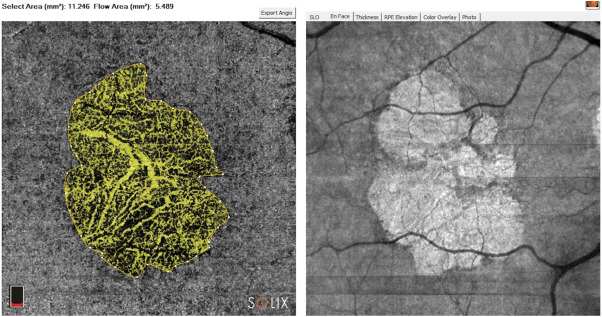
Figure 1. OCTA and en face OCT images of an eye with geographic atrophy associated with dry AMD. The delineation of the atrophic area was used to track its progression during the study.
Study intervention
After screening, the patients underwent vitrectomy and subretinal application of CB-PRP.
The product was supplied by the umbilical cord blood bank of the Fondazione Policlinico Universitario Agostino Gemelli IRCCS.
The units used came from donations originally intended for the cord blood bank. Only samples that met the health criteria and tested negative for infectious agents were considered.
Tests were performed for:
human immunodeficiency virus;
hepatitis B;
hepatitis C;
syphilis;
microbiological contamination.
The CB-PRP used in the study was made from a pool of 15 units.
Pooling the units was intended to reduce natural variations in growth factor concentration between different donors.
After processing, the material was divided into sterile 1 ml aliquots and stored at –80 °C until use.
All preparation was carried out using closed systems and sterile connections.
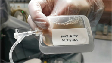
Figure 2. Sterile 1 ml aliquot of CB-PRP thawed and prepared for application.
Surgical procedure
Surgery was performed with 23 G pars plana vitrectomy and a high-speed system.
All procedures used the Constellation Vision system.
Patients received peribulbar anesthesia before the operation.
During the vitrectomy:
three valved cannulas were placed;
the posterior hyaloid membrane was removed when necessary;
a 41-gauge subretinal needle was introduced;
approximately 0.5 ml of CB-PRP was applied into the subretinal space;
the application site was chosen in the inferonasal quadrant;
peripheral vitrectomy was performed;
peripheral lesions, when identified, were treated;
at the end, a fluid-air exchange was performed.
After surgery, patients received specific positioning instructions for the first few days.
The inferonasal quadrant was chosen to reduce the risk of injury to the atrophic macular area during formation of the subretinal bleb.
Ophthalmological assessments
All patients underwent a complete ophthalmological examination at baseline and during follow-up.
The examinations included:
visual acuity on the ETDRS chart;
anterior segment biomicroscopy;
direct and indirect ophthalmoscopy;
intraocular pressure;
OCT;
OCTA;
focal electroretinogram;
flicker visual evoked potential.
The areas of atrophy were calculated by two independent specialists.
The demarcation was performed with the aid of semi-automated software, and agreement between the graders was considered adequate when Cohen's coefficient exceeded 0.9.
Measurements were obtained in both the treated eyes and the control eyes.
To increase safety, the first five patients were operated on with a minimum interval of 20 days between procedures.
Statistical analysis
Data were analyzed using SPSS 26 and GraphPad Prism 8.
Quantitative variables were presented as mean and standard deviation.
Repeated-measures analysis of variance was used to compare:
treated and untreated eyes;
different follow-up time points;
interaction between treatment and time.
The following were assessed:
visual acuity;
central macular thickness;
area of atrophy.
Tukey's test was used for post hoc comparisons.
For the electroretinogram and visual evoked potential results, the paired-samples t-test was used.
P values below 0.05 were considered statistically significant.
Results
The mean age of the participants at the time of surgery was 74.61 years.
Visual acuity
In the treated group, mean visual acuity showed an initial improvement during the first months, followed by a return to values close to those observed before the procedure at the end of 12 months.
In the untreated eyes, there was a progressive reduction in the mean number of ETDRS letters during follow-up.
The overall statistical analysis did not show a significant difference attributable to the treatment.
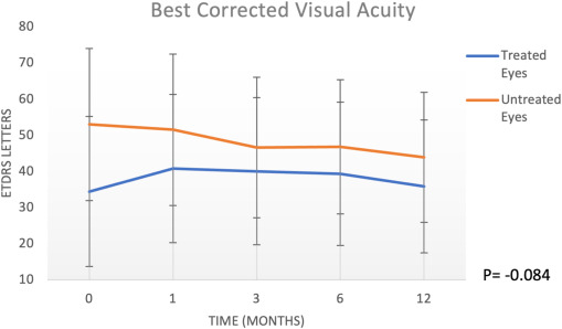
Figure 3. Progression of best-corrected visual acuity in treated and untreated eyes over 12 months. The bars represent the standard deviation.
Central macular thickness
Central macular thickness showed a gradual reduction in both groups.
At the end of 12 months, there was no significant difference between treated and untreated eyes.
However, at six months, post hoc analysis identified lower macular thickness in the control group.
This finding may suggest a possible temporary preservation of the macular structure in the treated eyes, but it is not sufficient to establish efficacy.
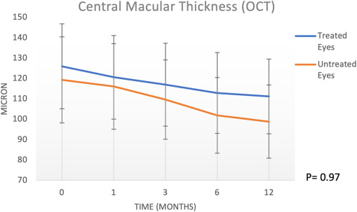
Figure 4. Central macular thickness of treated and untreated eyes at 1, 3, 6 and 12 months.
Progression of the area of atrophy
Agreement between the two specialists responsible for the measurements was greater than 90%.
The area of atrophy increased in both groups over the 12 months.
In the treated eyes, the mean area progressed from 15.16 mm² at baseline to 17.35 mm² at the end of follow-up.
In the untreated eyes, it went from 8.47 mm² to 11.59 mm².
The researchers observed a possible trend toward slower progression in the treated eyes at some early time points, especially between the first and third months.
However, since the treated eyes already had more advanced disease at baseline, this difference should be interpreted with caution.
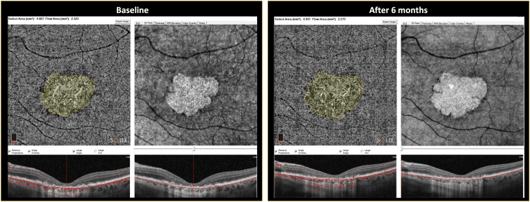
Figure 5. Semi-automated assessment of the area of geographic atrophy on en face OCT and OCT angiography images, comparing baseline with the sixth month.
Table 1 — Progression of the main parameters
Treated eyes
Parameter | Baseline | 1 month | 3 months | 6 months | 12 months | P value |
Visual acuity — ETDRS letters | 34,46 ± 20,8 | 40,84 ± 20,52 | 40,07 ± 20,34 | 39,38 ± 19,84 | 35,84 ± 18,38 | 0,06 |
Central macular thickness — µm | 120,54 ± 52,16 | 119,31 ± 50,91 | 118 ± 49,99 | 117,46 ± 53,91 | 111,23 ± 52,37 | 0,03 |
Area of atrophy — mm² | 15,16 ± 9,92 | 15,18 ± 9,92 | 15,74 ± 9,83 | 16,98 ± 9,88 | 17,35 ± 9,78 | < 0,0001 |
Square-root-transformed annual progression | 0,2709 | — | — | — | — | — |
Untreated eyes
Parameter | Baseline | 1 month | 3 months | 6 months | 12 months | P value |
Visual acuity — ETDRS letters | 53 ± 21,1 | 51,54 ± 20,99 | 46,62 ± 19,47 | 46,85 ± 18,58 | 43,92 ± 17,97 | 0,02 |
Central macular thickness — µm | 119,31 ± 58,52 | 118,92 ± 56,16 | 117,46 ± 58,52 | 107,07 ± 54,97 | 98,85 ± 54,55 | < 0,001 |
Area of atrophy — mm² | 8,47 ± 4,52 | 9,47 ± 4,85 | 10,00 ± 4,86 | 11,17 ± 4,91 | 11,59 ± 4,75 | < 0,0001 |
Square-root-transformed annual progression | 0,4946 | — | — | — | — | — |
Functional tests
The mean amplitudes of the focal electroretinogram and the visual evoked potential decreased at six months in both the treated eyes and the control eyes.
At 12 months, the behavior of the functional responses no longer showed the same pattern observed at the sixth month.
The authors consider the possibility that transient changes may be related to the temporary action of the growth factors.
However, since similar changes also occurred in the untreated eyes, it was not possible to establish a direct relationship with CB-PRP.
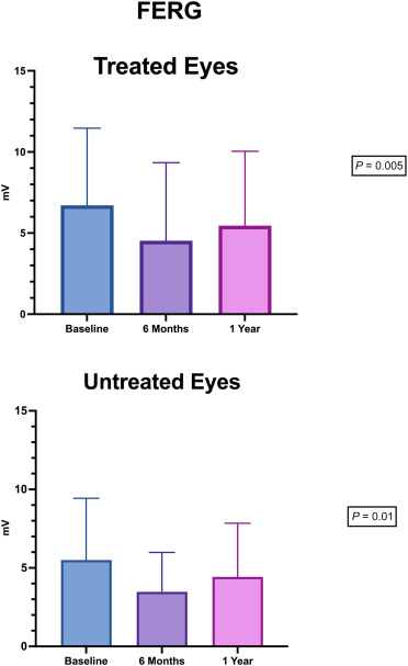
Figure 6. Focal electroretinogram amplitude in treated and untreated eyes during follow-up.
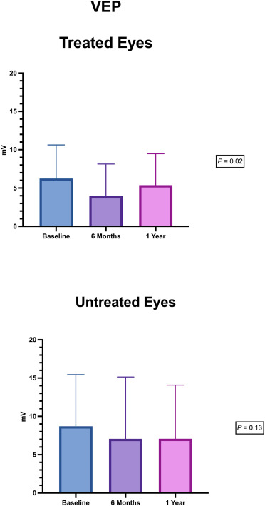
Suggested caption:Figure 7. Flicker visual evoked potential amplitude in treated and untreated eyes.
Safety
None of the patients had complications directly related to the subretinal application of CB-PRP during the 12 months.
The following were not observed:
endophthalmitis;
uveitis;
relevant inflammation;
retinal detachment;
product-related retinal lesions;
adverse events requiring discontinuation of the study.
This result supports the main objective of the research: to demonstrate the feasibility and initial safety of the technique.
Discussion
Research on geographic atrophy has been advancing on several fronts.
For several years, anti-inflammatory agents and complement system inhibitors were evaluated as alternatives to reduce disease progression.
Several substances did not achieve the expected results in clinical studies. Others, such as pegcetacoplan and avacincaptad pegol, demonstrated the ability to slow the growth rate of the atrophic area.
These therapies represented an important advance, but they do not recover tissue that has already been lost and depend on repeated intravitreal injections.
In this context, neuroprotective strategies continue to attract interest.
CB-PRP contains a wide variety of growth factors and biological mediators, including:
platelet-derived growth factor;
insulin-like growth factor;
epidermal growth factor;
hepatocyte growth factor;
transforming growth factor;
fibroblast growth factor;
vascular endothelial growth factor;
nerve growth factor;
various interleukins.
These substances take part in processes related to wound healing, regeneration, cell survival and modulation of the inflammatory response.
In ophthalmology, umbilical cord blood derivatives are already used or investigated mainly in ocular surface diseases.
There are reports of their use in:
corneal ulcers;
dry eye syndrome;
neurotrophic keratitis;
chemical injuries;
persistent epithelial defects;
recovery after corneal procedures.
The current study broadens this discussion by investigating subretinal administration.
The results showed that a single injection was well tolerated over 12 months.
A possible signal of slower atrophy progression was also identified at some early time points. However, this finding cannot be considered a demonstration of efficacy.
The reasons include:
small number of participants;
lack of randomization;
baseline differences between the treated eyes and the controls;
choice of the more affected eye to receive the treatment;
inability to fully separate the effect of CB-PRP from the effect of the vitrectomy itself.
The researchers also observed structural changes at the margins of the atrophic area, interpreted as a possible remodeling of the retinal pigment epithelium.
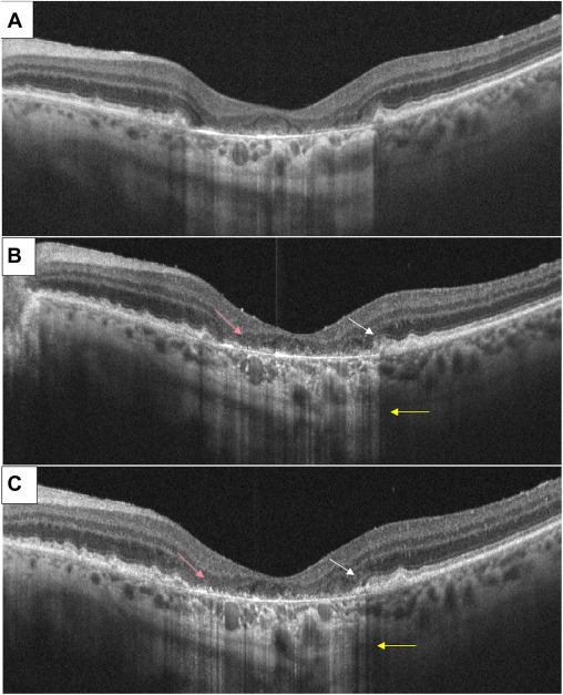
Figure 8. Structural OCT before the application and after three and six months. The images show changes at the margin of the atrophic area, interpreted by the authors as possible remodeling of the retinal pigment epithelium.
The subretinal route was chosen to overcome the inner blood-retinal barrier and place the biological factors close to the target tissue.
This type of administration is already used in some gene and cell therapies.
Despite the safety observed, subretinal application requires a surgical procedure and does not lend itself to frequent repetition of the treatment.
For this reason, the authors suggest that other routes of administration be evaluated in future research.
Comparison with complement inhibitor treatments
The study makes an exploratory comparison between the atrophy progression observed with CB-PRP and published data on complement inhibitors.
However, these comparisons must be interpreted with great caution because the study designs are different.
Pegcetacoplan and avacincaptad pegol were evaluated in larger, controlled trials with periodic intravitreal injections.
In the Si.Cord study, only a single subretinal application was performed in a small group without randomization.
Therefore, it is not possible to conclude that CB-PRP is equivalent, superior or inferior to these treatments.
Visual acuity
In the treated eyes, visual acuity showed an initial improvement and then returned to levels close to baseline values.
The authors consider that part of this initial improvement may be related to the removal of the vitreous during surgery.
A vitrectomy can make the ocular media clearer and produce a small visual gain in some patients.
In the control eyes, vision declined over 12 months, consistent with the natural history of geographic atrophy.
Structural changes
Central macular thickness did not show a significant difference between the groups at the end of the study.
The difference observed at six months may represent a possible transient effect, but it needs to be confirmed in a larger sample.
Regarding the area of atrophy, the treated eyes showed slower growth at some early time points.
However, since these eyes already had larger lesions and more advanced disease, their rate of expansion could naturally be different.
This factor makes it difficult to interpret the results.
Electrophysiological results
The functional tests revealed a reduction in amplitude at six months.
The authors hypothesize that a temporary modulation of retinal sensitivity could be associated with the activity of the growth factors.
However, since the same phenomenon also occurred in the untreated eyes, there is not enough evidence to attribute it to CB-PRP.
Another hypothesis is that the biological effect of the product has a duration limited by the half-life of the growth factors.
Studies with longer follow-up and different assessment intervals will be needed to clarify this point.
Limitations
The study has important limitations:
it was conducted at a single center;
there was no randomization;
the study was open-label;
only 13 patients took part;
the treated eyes already had more advanced disease;
there were baseline differences in visual acuity and lesion size between the groups;
follow-up was limited to 12 months;
only one application was performed;
the subretinal route makes repeated applications difficult;
the vitrectomy may have influenced some visual outcomes.
These limitations prevent definitive conclusions about efficacy.
Strengths
One of the main strengths was the standardization of the CB-PRP.
The product used resulted from pooling 15 umbilical cord blood units.
This process reduced variability between donors and allowed all aliquots to have a more homogeneous biological profile.
Another strength was the comparative follow-up of the treated eye and the fellow eye in each patient.
Assessment by two independent specialists also increased the reliability of the measurements of the area of atrophy.
Final considerations
The Si.Cord Study provided the first clinical evidence that a subretinal injection of CB-PRP can be performed safely in patients with geographic atrophy associated with dry AMD.
Over 12 months, no relevant complications related to the product or the procedure were observed.
The results also showed some exploratory signals that may justify further research, including:
possible slower rate of progression of the atrophic area in certain periods;
possible temporary preservation of macular thickness;
structural changes at the margins of the atrophy.
These findings, however, do not prove therapeutic benefit.
Larger, randomized, controlled studies with prolonged follow-up will be needed to determine whether CB-PRP can play a real role in retinal neuroprotection.
It will also be important to investigate routes of administration that are less invasive and better suited to repeated treatments.
Acknowledgments
The authors acknowledged the support of the Ministero della Salute – Ricerca Corrente 2023.
Supplementary data
Video of the procedure
Suggested caption:Video 1. 23 G vitrectomy followed by subretinal application of CB-PRP with a 41-gauge needle in the nasal quadrant of the retina.
References
The scientific references should be kept as in the original article, preserving the numbering used throughout the text.
For publication on an institutional page or blog, it is recommended to include at the end:
the full reference of the main article;
the DOI;
the scientific references cited;
a notice that the content is for informational purposes;
the guidance that any treatment depends on an individualized medical assessment.
Important notice
This content presents the results of a pilot study with a small number of participants.
Subretinal CB-PRP remains an experimental approach for geographic atrophy and should not be presented as a proven treatment, a cure or a substitute for authorized therapies.
Any procedure should be recommended exclusively by an ophthalmolo


Comments